Esthetic Provisional Crowns Efficiently Prepared Without External Matrix
Kanokraj Srisukho, DDS, MS; Mary A. Baechle, DDS; and Ronald E. Kerby, DMD
Preparing a tooth to receive a full-coverage crown requires many skills to be mastered. From achieving profound anesthesia to tooth preparation and obtaining the final impression, one must perform all of these procedures accurately and efficiently. One of the steps in this process that is vital to preserving tooth form while the crown is being made is the fabrication of a functional and esthetic interim restoration.
Interim restorations can be created through either custom fabrication or made from preformed materials.1 A custom interim restoration is created by constructing a replica of the external surface of the tooth (or teeth) to be prepared. This replica is a matrix or template that can be made from a number of different materials and in a variety of ways. One can create this matrix through either direct means, ie, intraorally, or indirect methods via a model of the patient's teeth or a modified diagnostic model.2 Once this matrix has been created it can be filled with the desired interim material of choice and applied to the prepared tooth intraorally. After the material has polymerized, the matrix and set material are removed from the mouth. The set material is then separated from the matrix, trimmed, and polished. A custom interim restoration has now been produced with an internal surface custom fit to the preparation, and an external surface that is a reproduction of a diagnostic wax-up or the original pre-prepared tooth.
Likewise, pre-fabricated interim restorations are also available in various forms.2 The basic form consists of a shell of plastic, cellulose acetate, or metal of the appropriate size which is adjusted to fit properly over the prepared tooth.1 The shell is frequently relined and then cemented directly onto the prepared tooth.1
Recently, a preformed malleable bis-acryl interim material, the Protemp Crown Temporization Material, was introduced by 3M ESPE (St. Paul, MN).
PRODUCT DESCRIPTION
Unlike other esthetic interim materials that are designed for short-term provisionalization and are chemically polymerized, the Protemp Crown Temporization Material is composed of a light-polymerized dimethacrylate Bis-GMA-based composite. It has a self-supporting, malleable consistency before light activation, which allows it to be trimmed, placed, and shaped to custom fit cuspid, bicuspid, or molar tooth preparations.
The Protemp Crown Temporization Material comes packaged either in a Trial or Promotional Kit. The Trial Kit includes temporary crowns for both bicuspid and molar crowns, crown size tools, instructions, a technique guide, and a CD. The Promotional Kit is larger and offers additional materials such as cuspid temporary crowns, a crown drawer with labels, and a technique DVD and booklet.
CASE REPORT
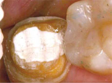
A 40-year-old woman presented for a crown preparation of the mandibular left second molar. After profound anesthesia was obtained, the tooth was prepared for a full-coverage, all-ceramic alumina Procera® Crown (Nobel Biocare USA, LLC, Yorba Linda, CA), with a 90° shoulder circumferential finishing line (Figure 1). Ultrapak Cord #1 (Ultradent Products, Inc, South Jordan, UT) impregnated with Hemodent (Dentalcompare, South San Francisco, CA) was placed in the gingival sulcus to displace the gingival tissue. After the final impression and bite registration were taken, the clinician began making the provisional restoration using the Protemp Crown Temporization Material.
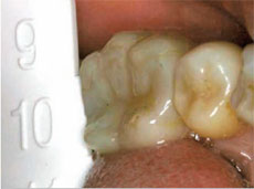
To determine the proper Protemp crown size, the mesio-distal width of the space available for the interim restoration was measured using the Crown Size Tool that is included in the kit (Figure 2). The Crown Size Tool is a measuring device that is tapered in decreasing increments from 12 mm to 7 mm in width. The tool was inserted on the occlusal surface of the crown preparation until it was lightly touching the distal surface of the tooth immediately mesial to it, at the level of the interproximal contact area. (When using the Crown Size Tool on a tooth with teeth both mesial and distal to it, one would insert the tool until it is slightly wedged between the teeth immediately adjacent to the preparation.) This width was then used to look up the corresponding Protemp crown size on the Size Selection Chart. Additionally, the occluso-gingival height of the adjacent crown was measured with a periodontal probe so as to help estimate the occluso-gingival height of the provisional restoration (Figure 3). It is important that the appropriate-sized crown is selected before opening the individually packaged Protemp crown. The reasons for this are that the crowns are manufactured for one-time use, are light-sensitive, and cannot be disinfected once they are removed from their package.
A package containing the preformed provisional crown was then opened and the thin film that comes attached to the cervical portion of the crown was peeled off (Figure 4 and Figure 5). According to the manufacturer, this film is part of the manufacturing process that does not serve any application purposes, such as light protection or pre-polymerization prevention. The malleable interim was then trimmed with crown-and-bridge scissors cervically to match the contour of the preparation margin and the occlusal height measured previously by the periodontal probe (Figure 6).
After trimming, the crown was seated on the moist, prepared tooth to verify the height. Then the crown was adapted to all of the prepared tooth surfaces and finish line with slight finger pressure, while also establishing interproximal contacts (only one to establish). The patient was then asked to gently close to establish the occlusion (Figure 7). While the patient was biting in occlusion, the interim margins facially accessible were adapted to the tooth with light finger pressure and an 8A Composite Instrument (Hu-Friedy, Chicago, IL). While the patient was still closed the facial aspect of the interim restoration was light-cured for 2 to 3 seconds. It is recommended not to light-cure longer than a few seconds for this initial tack-cure to allow for removal and adjustment of the provisional before final cementation.
The patient then opened her mouth and a ball-burnisher instrument (27/29 Burnisher, Hu-Friedy) was used to adapt the occlusal surface of the interim crown to the prepared occlusal surface (Figure 8). The lingual margin was then adapted to the tooth using the 8A instrument, while applying finger pressure to the buccal surface so as to prevent the crown from dislodging. The lingual and occlusal surfaces were then tack-cured for 2 to 3 seconds each. After carefully removing the partially cured interim crown from the patient's mouth, the crown was light-cured for a total of 60 seconds. The crown was placed back into the patient's mouth to check the fit and occlusion and to make any final adjustments before cementation. The crown was then removed from the patient's mouth to be finished and polished with composite finishing burs followed by Sof-Lex Contouring and Polishing Discs (3M ESPE) (Figure 9 and Figure 10).
The interim crown was now ready for final cementation. The prepared tooth was cleaned and lightly air-dried. Zone Temporary Cement (Prestige Dental Products Ltd, Yorkshire, UK) was mixed and applied to the internal surface of the crown, and care was taken to prevent air entrapment during mixing and seating of the interim. The final occlusion was then checked, and all residual set cement removed and cleaned off the interim restoration and tooth (Figure 11 and Figure 12).
CONCLUSION
The authors found that the prefabricated Protemp Crown Temporization Material produced an esthetic provisional restoration with good anatomical contours and occlusion. Good marginal adaptation was also achieved as a result of the malleability of the material before light polymerization.
The authors also found the interim restoration to fit very accurately so as to require only minimal adjustment with composite burs. Relining with composite resin was not required. If, however, there are any discrepancies in the anatomical contour or margins of a Protemp interim, they could be easily adjusted by adding a flowable composite resin where needed, and then trimmed with composite finishing burs. In addition, the authors observed that the provisional was very easy to trim and did not catch or grab the blades of the finishing burs. Trimming and polishing the crown produced an overall smooth surface, especially at the margins. Although the authors chose finishing burs and discs for final polishing, one may choose to use pumice and polishing compound instead.3-5
Because it is not polymethyl- or ethylmethacrylate-based, Protemp Crown Temporization Material did not create an unpleasant taste or odor throughout the fabrication, trimming, or cementation process, nor did it produce a highly exothermic setting reaction. This type of reaction would have required early removal of the interim restoration from the prepared tooth to prevent damage to the pulp. This may also have resulted in an unsatisfactory fit as a result of the subsequent high amount of polymerization shrinkage.6,7
The authors observed that the chair time consumed to make the Protemp provisional was less than that used to fabricate a chemically polymerized, tooth-colored provisional restoration. This can be quite advantageous in situations where patients demand esthetic posterior interim restorations, and there is a time constraint in which to fabricate an interim restoration. It is also helpful in situations where a provisional must be fabricated when a template of the pre-prepared tooth is not available or when there is little time to make such a template. Therefore, the authors find Protemp Crown Temporization Material to be a good option for dentists to choose when fabricating an esthetic custom interim restoration for cuspids and posterior teeth.
ACKNOWLEDGMENTS
The authors would like to thank Dr. Robert Rashid for converting the photographs for publication.
References
1. Burns DR, Beck DA, and Nelson SK. A review of selected dental literature on contemporary provisional fixed prosthodontic treatment: Report of the committee on research in fixed prosthodontics of the academy of fixed prosthodontics. J Prosthet Dent. 2003;90: 5:474-497.2. Rosenstiel SF, Land MF, Fujimoto J. Contemporary Fixed Prosthodontics. 4th ed. St. Louis: Mosby Elsevier; 2006:466-504.
3. Sen D, Goller G, Issever H. The effect of two polishing pastes on the surface roughness of bis-acryl composite and methacrylate-based resins. J Prosthet Dent. 2002;88: 527-532.
4. Maalhagh-Fard A, Wagner WC, Pink FE, Neme AM. Evaluation of surface finish and polish of eight provisional restorative materials using acrylic bur and abrasive disk with and without pumice. Oper Dent. 2003;28: 734-739.
5. Shillingburg HT Jr, Hobo S, Whitsett LD, et al. Fundamentals of Fixed Prosthodontics. 3rd ed. London: Quintessence; 1997:225-256.
6. Tjan AH, Grant BE, Godfrey MF 3rd. Temperature rise in the pulp chamber during fabrication of provisional crowns. J Prosthet Dent. 1989;62:622-626.
7. Castelnuovo J, Tjan AH. Temperature rise in pulpal chamber during fabrication of provisional resinous crowns. J Prosthet Dent. 1997;78:441-446.
 |  | |
| Figure 1 Tooth No. 18 prepared for a Procera all-ceramic crown. | Figure 2 Crown size tool placed on the occlusal surface of the prepared tooth. | |
 |  | |
| Figure 3 Periodontal probe used to measure the occluso-gingival height. | Figure 4 A thin film comes attached to the cervical portion of the Protemp crown. | |
 |  | |
| Figure 5 Peeling off the thin film. | Figure 6 Trimming the Protemp crown with crown-and-bridge scissors. | |
 |  | |
| Figure 7 Occlusion established by the patient?s bite and articulating paper. | Figure 8 Stabilizing the provisional crown with a ball-burnisher and adapting the material cervically with an 8A composite instrument. | |
 |  | |
| Figure 9 Adjusting occlusion with a composite finishing bur. | Figure 10 Finishing and polishing with a Sof- Lex Contouring and Polishing Disc. | |
 |  | |
| Figure 11 Occlusal view of the Protemp Crown after trimming and polishing. | Figure 12 Protemp interim restoration cemented to the tooth. |
| ABOUT THE AUTORS |
 Kanokraj Srisukho, DDS, MS Kanokraj Srisukho, DDS, MSAssistant Professor of Clinical Dentistry Section of Restorative and Prosthetic Dentistry The Ohio State University Health Sciences Center College of Dentistry Columbus, Ohio |
 Mary A. Baechle, DDS Mary A. Baechle, DDSAssistant Professor of Clinical Dentistry Section of Primary Care The Ohio State University Health Sciences Center College of Dentistry Columbus, Ohio |
 Ronald E. Kerby, DMD Ronald E. Kerby, DMDAssociate Professor Section of Restorative and Prosthetic Dentistry The Ohio State University Health Sciences Center College of Dentistry Columbus, Ohio |

