Conservative and Predictable Implant Therapy
Abstract
The practice of general dentistry has become increasingly demanding from the standpoints of diagnosis, treatment planning, ensuring high-quality care, and assuming the responsibility for the overall management of patient care. The diagnostic and treatment planning skills of the general dentist are of great importance. Implant dentistry requires advanced knowledge of the various dental disciplines and a planned sequence of therapy with various stages of evaluation. This case demonstrates the organization and implementation of total patient care; pre-prosthetic and strategic extraction of the maxillary first molar; evaluation and healing of the extraction socket; timing the placement of the implant; choosing the abutment and final restoration of choice; occlusal considerations; and maintenance therapy.
Implant dentistry has been integrated into the various dental disciplines and has significantly changed the scope of dentistry for both the generalist and specialist. The innovations and advances associated with bone regeneration and implantation have taken the practice of dentistry into a new and exciting era, adding a new armamentarium to clinical practice and each dental discipline.
The purpose of this case report is to present total patient care, from replacement of a maxillary first molar in the emergent treatment planning phase of therapy to the placement of the final implant and maintenance thereof. The patient care presented includes periodontics, oral surgery, implant dentistry, occlusion and restorative dentistry.
Case Report
A 34-year-old female patient presented with extreme pain in the upper right quadrant. The patient had a history of sensitivity when eating for the past month and severe pain in the past 3 days. Medical history was non-contributory. The patient admitted to smoking one pack of cigarettes and drinking two alcoholic beverages per day. Her last dental visit was 2 years prior. She isolated the pain on chewing to the right side of her face and radiating to her temple. There was sensitivity to percussion on teeth Nos. 2 and 3.
She had generalized wear, soft tissue inflammation, light calculus and stain, as well as generalized 2 mm to 4 mm pocket depths, except for tooth No. 3. Tooth No. 3 had a mesial and distal class III furcation and 7 mm to 8 mm probing depths along the mesial and palatal roots. These probing depths extended between all three roots, however the buccal probing was 2 mm. Therefore the periodontal lesion in the interradicular space of the furcation of tooth No. 3 was expansive. The extent of the deformity ruled out saving the two buccal roots. There was generalized 1+ degree mobility.
Radiographs revealed widened periodontal ligament (PDL) of the maxillary molars associated with occlusal traumatism and severe bone loss in the furcation area of tooth No. 3. The crestal bone of the remaining dentition was intact. There was generalized 1+ degree mobility created by the fractured root between the three roots of the maxillary first molar (Figure 1, Figure 2 and Figure 3).
Diagnosis
The diagnosis was established as primary occlusal traumatism with gingivitis and localized periodontitis associated with tooth No. 3, causing a vertical root fracture that resulted due to occlusal trauma from nocturnal grinding. The etiology for the gingivitis included: cigarette smoking, light calculus, poor home care, and neglect.
Formulating the Treatment Plan
The patient was presented the following options for tooth replacement: implant, 3-unit fixed bridge, Maryland bridge, removable partial denture, and no replacement. The ideal treatment plan for this molar was extraction followed by implant restoration, while the remaining dentition required periodontal and occlusal therapy. The therapeutics entailed a series of steps building on each other to reach the final goal.
- Systemic antibiotic therapy and extraction of tooth No. 3
- Full-mouth scaling and home care instruction
- Occlusal adjustment by selective grinding and night guard therapy
- Evaluate socket healing and soft tissue response to scaling
- Place implant in area of tooth No. 3 (bone graft if needed), 8 to 16 weeks postoperative
- Second stage surgery 12 to 16 weeks after implant placement; evaluate bone, soft tissue, implant stability; fabricate temporary and final impression of implant coping
- Place the final restoration 2 to 4 weeks later; retrofit the night guard
- Maintenance therapy at 3-month intervals, focusing on occlusion, periodontium, and implant restoration
This is a simple, organized treatment sequence with many painstaking steps, complicated by cigarette smoking and nocturnal grinding. Throughout treatment the author monitored the bone, soft tissue, and occlusion, and will continue to do so during maintenance therapy.
Tooth Preparation for Sectioning Roots
The first step in sectioning this multi-rooted tooth was to remove any crowns or proximal surfaces to gain access to furcation openings. Decornication of the tooth was performed with a surgical carbide in a highspeed handpiece at the gingival margin. A thin diamond stone was placed in the furcation opening, moved through the coronal aspect of the tooth, and connected to the other furcation opening, separating the tooth into individual roots. Following the complete separation of the roots, the interradicular surfaces were prepared to gain access to the individual roots. The distal root was removed first (Figure 4); then the mesial root was removed with a periotome or thin elevator on the proximal surfaces. The palatal root was the last to be removed and was moved to the buccal aspect of the socket.
Strategic Extraction
The strategic extraction of the tooth begins with examination of the radiographs and performing periodontal probing to consider the extent of the lesion and the remaining osseous walls. Clinically, the soft tissue character, purulunce, and sulcular fluid, as well as the osseous topography, should be evaluated.1 This information is important in planning the strategic extraction to ensure the sound osseous walls are protected from the trauma of the extraction.
Evaluation and Healing of Extraction Socket
In this case, there were five osseous walls intact: mesial, distal, buccal, palatal, and the base (toward the sinus).2 The interradicular osseous septum had been destroyed by the disease process. The disease granulation tissue was removed and immediate clotting was observed. The patient’s smoking was a deterrent to bone grafting. It was predicted that natural healing would occur, but would be delayed (due to smoking and the extent of the lesion) for implant placement in 12 to 16 weeks. Healing therefore would be evaluated radiographically and clinically at 4-week intervals.
When the osseous topography has five intact walls and the patient is healthy, healing of the extraction socket is predictable. The author recommends implant placement at the 6-to-8-week interval (delayed immediate placement). The concept of preprosthetic strategic extraction and evaluation allows for maximum preservation of the osseous structures and predictable healing patterns in the posterior maxilla.3
In this case, radiographs at 4-week intervals revealed that the tissue was slow to mature because of cigarette smoking. On day 112 postextraction, the implant was inserted (Figure 5). At that time, the bone was well matured and the soft tissue was healthy.4
At the time of implant placement, the character of the bone and the stability of the implant can be evaluated and the healing time determined. In this case, the implant was uncovered 12 weeks postinsertion.
Implant Placement
The GS II (Hiossen, www.hiossen.com), straight-bodied, internal-hexed, bone-level implant was placed. This implant was chosen because of its drilling armamentarium, self-tapping capacity, initial stability, ease of placement, bioaffinity, and reliability. The 11-degree Morse taper of the internal connection provides a stable connection to the superstructure. Employing this particular implant in the posterior maxilla provided increased body and surface area. The final restoration planned would be a one-piece screw-retained cast gold abutment and porcelain was fused to the restoration.
The patient was placed on a routine dose of antibiotics. A conservative flap procedure gained access to the healed extraction socket. The osseous crest was intact, as were the buccal and lingual walls. There was 10 mm of bone buccolingually, 10 mm mesiodistally, and 10 mm to 11 mm from crest to the floor of the sinus. There was interocclusal clearance of 8 mm to 9 mm. A 5-mm by 10-mm Hiossen straight-bodied GS II fixture was placed. The flap was repositioned and four gut-interrupted sutures were placed (Figure 6). The healing time for osseous integration was 12 weeks (day 84).
Second Stage Surgery and Coping Pickup
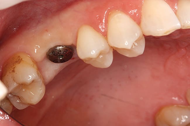
The second stage surgical procedure should be approached cautiously to enhance the soft tissue needs. There should be at least 3 mm to 4 mm of bound-down attached gingiva to protect the soft tissue surrounding the implant providing stable soft tissue long term. In this case, there was adequate soft tissue for the extraction of the tooth and placement of the implant in the posterior maxilla. However, the implant was partially exposed because of the patient’s smoking (Figure 7). A surgical punch incision was employed to completely expose the implant. Also at this time, the closed-tray engaging the implant coping was placed onto the fixture (Figure 8) and a full arch impression for the fabrication of the final restoration was made, as well as full-arch counter impression and bite records. A 5.5-mm by 3-mm healing abutment was placed at the gingival level (Figure 9). A temporary restoration would have been placed if necessary.
Final Restoration
A screw-retained, one-piece, engaging UCLA implant abutment with porcelain was the restoration of choice (Figure 10 and Figure 11). The advantage of this restoration is its simplicity and predictability. The abutment connection was an 11-degree Morse taper with an internal hex that geometrically locked with an internal hex. The implant abutment junction was medialized (platform switched) by the design of the GS II implant, which—along with the rigid abutment connection—distributes forces below the osseous crest and prevents crestal bone remodeling.
Occlusion
The implant must be protected from excessive and occlusal overload. The buccal cusp of the mandibular molar should be positioned to occlude in the central fossa of the maxillary restoration. The palatal cusp of the maxillary first molar must be narrow to diminish the cantilever effect of the implant and allow clearance in all protrusive and lateral protrusive movements. The maxillary buccal cusp should be modified to allow clearance during the Bennett shift and lateral movement on the working side.5
Maintenance Therapy
Maintenance therapy includes monitoring patient complaints, health, and general dental pathologic entities. Specifically, the implant restoration should be monitored for soft tissue health, the makeup of the osseous topography, and most importantly, the occlusion (the presence of excessive force). Over time, the dentition wears; the porcelain restoration and the rigid implant may become an occlusal interference. Therefore, the clinician should be prepared to do occlusal adjustment on routine maintenance visits. Maintenance therapy should be scheduled on 3-to-6-month intervals, depending on the extent of the restoration, the periodontal climate, and the patient’s susceptibility to disease (Figure 12). With the patient’s history of clenching nocturnally, use of a night time occlusal guard is strongly emphasized to prevent overloading of the implant and subsequent periodontal issues.
References
1. Casullo D. The integration of endodontics, periodontics and restorative dentistry in general practice. Part I. Diagnosis. Compend Contin Educ Gen Dent. 1980;1(2):137-147.
2. Goldman HM, Cohen DW. The infrabony pocket: classification and treatment. J Periodontol. 1958;29:272.
3. Schropp L, Wenzel A, Kostopoulos L, Karring T. Bone healing and soft tissue contour changes following single tooth extractions: A clinical and radiographic 12-month prospective study. Int J Periodontics Restorative Dent. 2003;23(4):313-323.
4. Cardaropoli G, Araújo M, Lindhe J. Dynamics of bone tissue formation in tooth extraction sites. An experimental study in dogs. J Clin Periodontol. 2003;30(5):809-818.
5. Abrams L. Occlusal adjustment of the natural dentition. Chicago, IL: Quintessence; 1986.
Disclosure
The author is a part-time instructor for Hiossen.
About the Author
*Clinical Associate Professor University of Pennsylvania
School of Dental Medicine
Philadelphia, Pennsylvania
Private Practitioner
Philadelphia, Pennsylvania


