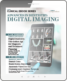
Advances in Dentistry: Digital Imaging
Wednesday, June 20, 2018
Commercial Supporter:

Volume 38 eBook 26
In this special eBook from Compendium, we offer two continuing education articles designed to keep the dental professional updated on how digital dental technology is replacing traditional workflows and becoming the new standard in dentistry. The first CE article describes the benefits of acquiring a digital replica of dentoalveolar structures accurately and non-invasively with several current systems, offering an immediate advantage over conventional impressions. The second CE article examines how intraoral cone-beam tomography (CBCT), otherwise known as volume imaging CT scan, provides 3D images of mandibular and maxillary structures. These highly accurate scans provide valuable diagnostic information to facilitate treatment planning for implant cases.. Download to earn 4 FREE CEU now!
FEATURED CONTENT
CE 1: Digital Dentistry: The Golden Age for Diagnosis and Treatment Planning
Radi Masri, DDS, MS, PhD; Carl F. Driscoll, DMD; Se Jong Kim, DMD; and William M. Wahl, DDS
This first CE article describes the benefits of acquiring a digital replica of dentoalveolar structures accurately and non-invasively with several current systems, offering an immediate advantage over conventional impressions. The authors discuss how current digital systems facilitate and enhance diagnosis and treatment planning, which as a result refines treatment options. For example, optimal scans of teeth, or diagnostic wax-ups, can be combined with 3D radiographic images using the existing teeth as natural fiduciary markers to advance treatment in implant dentistry.
Credits: 2 Self-Study CEU
Cost: $0
Provider: AEGIS Publications, LLC
CE 2: Dental Cone-Beam Scans: Important Anatomic Views for the Contemporary Implant Surgeon
Gary Greenstein, DDS, MS; Joseph R. Carpentieri, DDS; and John Cavallaro, DDS
This second CE article examines how intraoral cone-beam tomography (CBCT), otherwise known as volume imaging CT scan, provides 3D images of mandibular and maxillary structures. These highly accurate scans provide valuable diagnostic information to facilitate treatment planning for implant cases. This article serves as a primer on how to read and interpret CBCT cross-sectional views. Ultimately, correct interpretation of CBCT reformatted images improves the predictability of implant placement and helps avoid unnecessary complications.
Credits: 2 Self-Study CEU
Cost: $0
Provider: AEGIS Publications, LLC


