Don't miss a digital issue! Renew/subscribe for FREE today.
×
Compendium
October 2023
Volume 44, Issue 9
Peer-Reviewed

Smile Restoration of a Dental Anxiety Patient Using Same-Day Chairside CAD/CAM and Esthetic Zirconia

Lincoln R. Fantaski, DMD

Abstract: This clinical case study describes the smile restoration of a 33-year-old female patient who exhibited severe dental anxiety, multiple cavities, discoloration, and a fractured tooth. The patient's complex dental history and anxiety-driven treatment preferences posed challenges that were met with a minimally invasive strategy. The case involved meticulous diagnosis, leveraging advanced diagnostic wax-ups, and a comprehensive treatment plan. Esthetic zirconia material was utilized to enable the preservation of tooth structure while achieving visually appealing and functional outcomes, and CAD/CAM technology facilitated same-day restorations with superior marginal fit, aided by the precision of a digital intraoral scanner. The patient's dental anxiety strongly influenced the treatment approach, leading to the integration of smile enhancement alongside caries treatment. The use of a single cement and bond product simplified the final cementation process, while the versatility of the zirconia CAD/CAM material allowed for fabrication of various restoration types with minimal tooth reduction. This streamlined workflow expedited treatment, which was especially crucial for the anxious patient undergoing sedation.

Patients with a history of dental trauma often shy away from discussing their smile goals due to anxiety and a belief that improvement is unattainable.1,2 The advent of same-day chairside CAD/CAM technology and esthetic zirconia has revolutionized dental restorations, making remarkable smile transformations possible while preserving tooth structure.3-5 This case study presents the successful restoration of an adult patient with severe dental anxiety, multiple cavities, discoloration, and a fractured tooth using a minimally invasive approach and esthetic zirconia restorations. Utilizing the zirconia material would enable the preservation of tooth structure while allowing esthetic and functional outcomes to be achieved.6,7 The implementation of CAD/CAM dentistry facilitated same-day restorations with exceptional marginal fit, supported by precise digital intraoral scanning.8-10

Patient Presentation

The patient, a 33-year-old woman, presented with complaints of several cavities, tooth staining, and discoloration around old restorations (Figure 1). She also said she had significant dental anxiety. She reported a pain level of eight out of 10 in tooth No. 14, which had undergone previous endodontic treatment with suspected fracture. The patient's dental history included recurrent childhood cavities with unidentified sources. Because of her anxiety, she requested sedation for all dental procedures.

Clinical Findings

Upon examination, the following conditions were identified for each treated tooth: Nos. 2 through 5 each had secondary caries under a previous composite restoration, with No. 3 also having primary caries on the distal aspect, and Nos. 4 and 5 having the restorations covering 75% of the tooth structure. Nos. 6 through 12 and 15 each had secondary decay under a previous composite restoration, with No. 6 also having staining and hypocalcification on the buccal surface, No. 7 having deep decay and cold sensitivity, and No. 8 having the composite covering more than 75% of the tooth structure; No. 8 also was previously endodontically treated. Tooth No. 13 had secondary decay under the previous glass-ceramic crown and was also previously endodontically treated. Finally, No. 14 had endodontic failure and suspected fracture with a broken file in the palatal root, causing severe sensitivity to percussion and palpation; a periapical radiolucency was noted on the palatal root.

Diagnosis and Treatment Plan

The patient was diagnosed with severe/high caries risk, old and leaky composites, a fractured tooth with endodontic failure in tooth No. 14, secondary and primary caries, and wear from significant dental restorations leading to a collapsed vertical dimension of occlusion.

The treatment plan involved the following procedures: direct restoration of tooth No. 2, full cuspal coverage crowns for teeth Nos. 3 through 5, minimal-preparation veneers/crowns for teeth Nos. 6 through 11, cuspal coverage crowns for teeth Nos. 12 and 13, immediate implant placement for tooth No. 14 with a custom titanium abutment and zirconia crown on implant No. 14 following osseointegration, and an indirect onlay for tooth No. 15.

Treatment Approach

The dental team opted for a minimally invasive treatment approach, with the intention of preserving as much tooth structure as possible while achieving optimal outcomes. A multilayered esthetic zirconia material (KATANA Zirconia ONE, Kuraray, kuraraydental.com) was selected for its exceptional strength and impressive esthetics achievable with minimal preparation.11,12 The use of CAD/CAM technology (CEREC®, Dentsply Sirona, dentsplysirona.com) allowed for same-day restorations with tight marginal fit, aided by the accuracy of a digital intraoral scanner (Primescan, Dentsply Sirona).

The patient's severe dental anxiety influenced the treatment plan, leading to the decision to enhance her smile simultaneously with addressing the caries. Proper clinical and esthetic pre-planning, aided by lab-provided diagnostic wax-ups, facilitated successful results. The patient underwent the procedure under oral conscious sedation as requested.

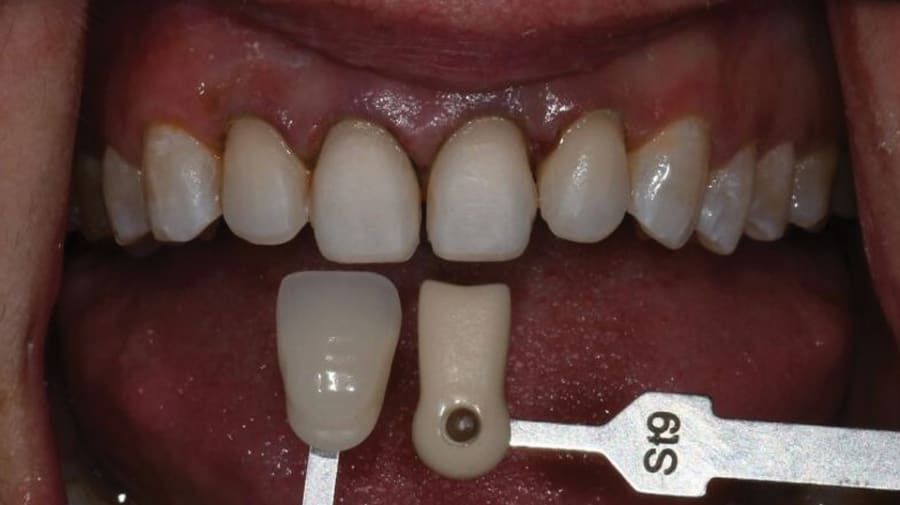
In addition to alleviating dental anxiety through sedation, the primary goals of the case were to optimize the patient's smile, utilize high-quality dental products and procedures to reduce secondary caries risk, eliminate pain and infection, and enhance the patient's confidence. The shade B1 of KATANA Zirconia ONE was chosen to enhance her smile while blending naturally with her mandibular arch, given the inherent esthetics of zirconia material.

Clinical Procedural Steps

Preparation went as follows: Under oral sedation, the patient was anesthetized, and a rubber dam was placed on the entire maxillary arch. Teeth were prepared using coarse diamond burs and carbides to remove old restorations, eliminate caries, and create an adequate surface roughness for micromechanical retention. For direct restorations, a total-etch technique was employed, and after etching, a cavity cleaner was used and dried before application of the bonding agent. For indirect restorations, the preparations were cleaned with cavity cleaner and dried before cementation.

Step 1: Teeth Nos. 2 through 5, 12, 13, and 15 were prepared. Old restorations were removed, caries was excavated, and preparations were refined. Core buildups and direct restorations were placed using a total-etch technique (CLEARFIL Universal Bond Quick, Kuraray) and a flowable composite (CLEARFIL MAJESTY ES Flow, Kuraray).

Step 2: A previously designed custom titanium abutment (Vulcan Custom Dental, vulcandental.com) was placed on the implant (BioHorizons, biohorizons.com) at the No. 14 position. Full-arch scans of the upper, lower, and bilateral buccal bite were taken (Primescan) (Figure 2). The diagnostic wax-up was scanned as a biocopy library in CEREC.

Step 3: Margins and copy lines were marked, and restorations were designed using the FOCC protocol (fissure height, occlusion, contours, contacts) with a biocopy of the wax-up (Figure 3). The designs were then sent to the mill (Primemill, Dentsply Sirona). Restorations Nos. 3 through 5 were milled and sintered first, followed by Nos. 12 through 14. After milling, each quadrant of restorations was sintered together.

Step 4: Posterior restorations were polished using the KATANA pre-sintering polishing protocol. The sprue was removed and polished using a carbide lab bur. The entire surface of each restoration was polished, first with a polishing brush, followed by fine and extra-fine polishing wheels (Meisinger, meisingerusa.com) (Figure 4). The restorations were then sintered in a high-speed furnace (SpeedFire, Dentsply Sirona).

Step 5: While restorations Nos. 12 through 14 were in the furnace, teeth Nos. 6 through 11 were prepared under a rubber dam. Caries was excavated, and significant caries was addressed on the "Class V" cementoenamel junction location on the buccal and interproximal under the contact points. Tooth No. 7 required and received root canal treatment, followed by bonding a fiber post into the post space. Teeth Nos. 8 and 9 were prepared for a full-coverage crown and veneer, respectively.

Step 6: Restorations for teeth Nos. 12 through 14 were seated. Preparations for teeth Nos. 6 through 11 were scanned, and a "locked in" temporary restoration was made (Luxatemp®, DMG America, dmg-america.com) using a putty mold of the diagnostic wax-up. The patient was awakened and dismissed for the day.

Step 7: Using the CAD/CAM software's biocopy feature (CEREC SW 5.2.8, Dentsply Sirona), the restorations for teeth Nos. 6 through 11 were designed and milled (Figure 5 and Figure 6). Characterization was added to the surface texture using a diamond and carbide burs (Figure 7). The restorations were then sintered and micro-etched or air-abraded using aluminum oxide. Minimal staining and glazing (CZR [CERABIEN ZR], Kuraray) were performed, and the restorations were fired using the glazing cycle on the SpeedFire furnace (Figure 8). The lingual aspect of each of these restorations was left unglazed to preserve the opposing dentition.

Step 8: The patient was reappointed under mild oral sedation. Temporary restorations were removed, and final crowns were tried in for marginal fit and contacts. Restorations were bonded two at a time (PANAVIA SA Cement Universal, Kuraray) starting with the central incisors and moving distal to the canines. The intaglio of each of the zirconia crowns was micro-etched prior to cementation. The cement was tack-cured, cleaned, and fully light-cured. The bite was adjusted, and the patient was dismissed.

Step 9: One week later, the patient returned for a follow-up appointment. The bite was checked, and final photographs were taken. The patient expressed overwhelming emotions, being amazed by the natural appearance and feel of her smile. The advancements in materials and technology and the staging of the case facilitated a successful smile transformation, resulting in a confident and satisfied patient (Figure 9 through Figure 11).

Discussion

During the final photograph session, the patient marveled at the natural beauty of her restored smile. Having battled cavities and dental anxiety throughout her life, she said she never imagined being proud of her smile. The restorations not only felt remarkably lifelike but also looked astonishingly natural, she acknowledged. The entire process seemed surprisingly easy to her, an achievement only made possible by the innovations in modern dental materials and technologies like same-day chairside CAD/CAM.

The versatility of the zirconia (KATANA Zirconia ONE) allowed for the incorporation of veneers, crowns, minimal-prep three-quarter or five-eighths crowns, and implant crowns with minimal tooth reduction, which was crucial, especially for a young patient. The built-in color gradient of the material enhanced the esthetics, eliminating the need for additional characterization and staining steps. Like natural teeth, crowns should exhibit natural color variations. Pre-polishing the zirconia proved time-saving and prevented the undesired "opal" appearance that can occur with overheated sintered zirconia. Moreover, using a single cement and bond product, without multiple priming steps for restoration and tooth structure, simplified the final cementation process. The tack-cure properties of the cement made cleanup effortless, and the range of color choices offered flexibility for achieving seamless harmony among all the restorations.

These material efficiencies and advancements facilitated the successful smile transformation, allowing the dental team to work swiftly on other aspects of the treatment while the patient rested comfortably and safely under sedation. Staging the case over two visits, with the patient returning to receive the anterior six units, and utilizing a porcelain system (CZR [CERABIEN ZR]) for hand characterization and glazing of the labial portion of the anterior restorations resulted in remarkably lifelike outcomes, uncommonly associated with "in-office" or same-day chairside dentistry. The natural gradient featured in the KATANA Zirconia ONE block made the staining process easier, with less staining needed to obtain a natural look as compared to monochromatic CAD/CAM blocks.

Conclusion

Through the combined use of same-day chairside CAD/CAM technology and esthetic zirconia, this case study demonstrates a successful and minimally invasive approach to restore a patient's smile, address dental anxiety, and improve the patient's overall dental health and confidence. The transformation of the patient's smile and her emotional response underscore the success of this approach. The multilayered zirconia material used in this case provides exceptional esthetics while offering wear tolerance and strength for long-term functional and cosmetic benefits. This case exemplifies the potential of modern dental advancements to achieve optimal outcomes for complex cases involving both clinical and psychological considerations.

Disclosure

This article was commercially supported by Kuraray.

About the Author

Lincoln R. Fantaski, DMD
Private Practice, Sandy Springs and Cumming, Georgia

References

1. De Stefano R. Psychological factors in dental patient care: odontophobia. Medicina (Kaunas). 2019;55(10):678.

2. Rafique S, Banerjee A, Fiske J. Management of the petrified dental patient. Dent Update. 2008;35(3):196-198.

3. Fasbinder DJ. The CEREC system: 25 years of chairside CAD/CAM dentistry. J Am Dent Assoc. 2010;141(suppl 2):3S-4S.

4. Sannino G, Germano F, Acruci L, et al. CEREC CAD/CAM chairside system. Oral Implantol (Rome). 2015;7(3):57-70.

5. Hehn S. The evolution of a chairside CAD/CAM system for dental restorations. Compend Contin Educ Dent. 2001;22(6 suppl):4-6.

6. Kasem AT, Sakrana AA, Ellayeh M, Ozcan M. Evaluation of zirconia and zirconia-reinforced glass ceramic systems fabricated for minimal invasive preparations using a novel standardization method. J Esthet Restor Dent. 2020;32(6):560-658.

7. Kasem AT, Elleyah M, Ozcan M, Sakrana AA. Three-year clinical evaluation of zirconia and zirconia-reinforced lithium silicate crowns with minimally invasive vertical preparation technique. Clin Oral Investig. 2023;27(4):1577-1588.

8. Akbar JH, Omar R, Al-Tarakmah Y. Marginal integrity of CAD/CAM ceramic crowns using two different finish line designs. Med Princ Pract. 2021;30(5):443-447.

9. Ng J, Ruse D, Wyatt C. A comparison of the marginal fit of crowns fabricated with digital and conventional methods. J Prosthet Dent. 2014;112(3):555-560.

10. Abduo J, Elseyoufi M. Accuracy of intraoral scanners: a systematic review of influencing factors. Eur J Prosthodont Restor Dent. 2018;26(3):101-121.

11. Inokoshi M, Liu H, Yoshihara K, et al. Layer characteristics in strength-gradient multilayered yttria-stabilized zirconia. Dent Mater. 2023;39(4):430-441.

12. Reyes AR, Dennison JB, Powers JM, et al. Translucency and flexural strength of translucent zirconia ceramics. J Prosthet Dent. 2023;129(4):644-649.

© 2024 Conexiant | Privacy Policy